


MUM1 (MRQ-8) Mouse Monoclonal Antibody
Specialties: Hematopathology
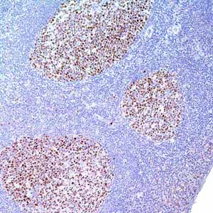
Anti-MUM1 antibody labels a 50kDa, multiple myeloma oncogen-1 (MUM1) protein. MUM1 is encoded by the MUM1/IRF-4 gene, which is mapped to 6q23-25 and identified as a myeloma-associated oncogene.1,2 It is a member of the interferon regulatory factor family of transcription factors and plays an important role in the regulation of gene expression in response to signaling by interferon and other cytokines. MUM1 positive cells express the protein in the nucleus in a diffuse and microgranular pattern. However, some positivity is also observed in the cytoplasm of MUM1-expressing cells. In normal/reactive lymphoid tissues, such as lymph node, this antibody stains plasma cells, some B-cells in the light zone of germinal centers, and a subset of T-cells (T-cells in germinal centers and interfollicular areas).1,2 MUM1 expression has been described in diffuse large B-cell lymphoma (DLBCL).4 Anti-MUM1 antibody can stain other B-cell lymphomas such as lymphoplasmacytic lymphoma, chronic lymphocytic leukemia, follicular lymphoma, marginal zone lymphoma, lymphoblastic lymphoma/leukemia, primary effusion lymphoma, DLBCL, Burkitt-like lymphoma, and classical Hodgkin lymphoma.3-5 However, the tumor cells in nodular lymphocyte predominant Hodgkin lymphoma are negative or only weakly positive.6 MUM1 is also expressed in plasma cell myeloma.7
- Falini B, et al. Antibody (MUM1P) detects expression of the MUM-1/IRF4 protein in a subset of germinal center B cells, plasma cells, and activated T cells. Blood. 2000; 95:2084-92.
- Grossman A, et al. Cloning of human lymphocyte-specific interferon regulatory factor (hLSIRF/hIRF4) and mapping of the gene to 6p23-25. Genomics. 1996; 37:229-33.
- Neresh KN. MUM1 expression dichotomizes follicular lymphoma into predominantly, MUM1-negative low grade and MUM1-positive high-grade subtypes. Haematologica. 2007; 92:267-8.
- Van Imhoff GW, et al. Prognostic impact of germinal center-associated protein and chromosomal breakpoints in poor-risk diffuse large B-cell lymphoma. J Clin Oncol. 2006; 24:4135-42.
- Gualco G, et al. MUM1/IRF4: A Review. Appl Immunohistochem Mol Morphol. 2010; 18:301-10.
- Carbone A, et al. Expression pattern of MUM1/IRF4 in the spectrum of pathology of Hodgkin’s disease. Br J Haematol. 2002; 117:366-72.
- Natkunam Y, et al. Analysis of MUM1/IRF4 protein expression using tissue microarrays and immunohistochemistry. Mod Pathol. 2001; 14:686-94.
Specifications
- Reactivity: paraffin
- Visualization: cytoplasmic, nuclear
- Control: tonsil
- Dilution Range: 1:100-1:500*
Package Inserts
IFU
- IVD Rev. 4.0 (CMC35821040)
Have a different keycode?
Click Here
Learn how to obtain your SDS
Ordering Information
For in vitro diagnostic (IVD) use in USA
| 0.1 mL concentrate | 358M-14 |
| 0.5 mL concentrate | 358M-15 |
| 1 mL concentrate | 358M-16 |
| 1 mL predilute | 358M-17 |
| 7 mL predilute | 358M-18 |
For in vitro diagnostic (IVD) use in Canada
| 0.1 mL concentrate | 358M-14 |
| 0.5 mL concentrate | 358M-15 |
| 1 mL concentrate | 358M-16 |
| 1 mL predilute | 358M-17 |
| 7 mL predilute | 358M-18 |
For in vitro diagnostic (IVD) use in Europe
| 0.1 mL concentrate | 358M-14 |
| 0.5 mL concentrate | 358M-15 |
| 1 mL concentrate | 358M-16 |
| 1 mL predilute | 358M-17 |
| 7 mL predilute | 358M-18 |
For research use only (RUO) in Japan
| 0.1 mL concentrate | 358M-14-RUO |
| 0.5 mL concentrate | 358M-15-RUO |
| 1 mL concentrate | 358M-16-RUO |
| 1 mL predilute | 358M-17-RUO |
| 7 mL predilute | 358M-18-RUO |
To request information on this product in additional countries, please click the button below.