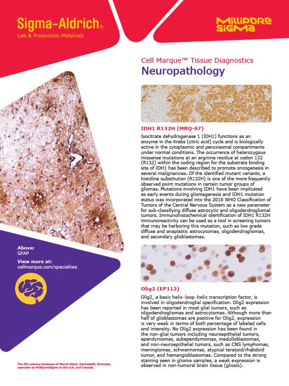
Glial Fibrillary Acidic Protein (EP672Y) Rabbit Monoclonal Primary Antibody
Specialties: Neuropathology
Detection of GFAP by immunohistochemistry (IHC) with Glial Fibrillary Acidic Protein (EP672Y) Rabbit Monoclonal Primary Antibody may be used to aid in the diagnosis of astrocytomas.
GFAP, or glial fibrillary acidic protein, is a 51-kDa intermediate filament protein that is a key element of the cytoskeleton structure of glial cells and helps maintaining their structural integrity as well as the integrity of the blood brain barrier. GFAP is normally expressed in glial cells such as astrocytes, ependymal cells, and choroid plexus, all of which are found only in the brain, as well as the pituitary gland, Muller cells of the retina, peripheral nerves, and stellate cells of the liver.1 Immunohistochemistry is positive in astrocytomas, which are primary brain tumors arising from astrocytes.2-4 Immunohistochemistry for GFAP shows a cytoplasmic pattern of staining.
In combination with other antibodies, this product is intended to be used as an aid in diagnosis.
- Redecker, P, and J Fechner."Immunohisto chemical study of cells positive for glial fibrillary acidic protein (GFAP) in the human pituitary gland, with special reference to folliculo-stellate cells."Histochemistry vol. 91,3 (1989): 227 34.
- Kimura, T et al."An immunocytochemical comparison of the glia-associated proteins glial fibrillary acidic protein (GFAP) and S-100 protein (S100P) in human brain tumors."Clinical neuropathology vol. 5,1 (1986): 21-7.
- Reifenberger, G et al."Differential expression of glial- and neuronal-associated antigens in human tumors of the central and peripheral nervous system."Acta neuropathologica vol. 74,2 (1987): 105-23.
- Rousseau, Audrey et al."Expression of oligodendroglial and astrocytic lineage markers in diffuse gliomas: use of YKL-40, ApoE, ASCL1, and NKX2-2."Journal of neuropathology and experimental neurology vol. 65,12 (2006): 1149-56.
- Nadji M, Morales AR. Immunoperoxidase, part I:the techniques and its pitfalls. Lab Med 1983;14:767
- Omata M, Liew CT, Ashcavai M, Peters RL. Non immunologic binding of horseradish peroxidase to hepatitis B surface antigen. A possible source of error in immunohistochemistry. Am J Clin Pathol. 1980 May;73(5):626-32. doi: 10.1093/ ajcp/73.5.626. PMID: 6155065.
Specifications
- Reactivity: paraffin
- Control: Brain (Cytoplasmic)
- Dilution Range: 1:25-1:100 *
Package Inserts
IFU
- IVD Rev. 6.0 (CMC25831060)
Have a different keycode?
Click Here
Learn how to obtain your SDS
Ordering Information
For in vitro diagnostic (IVD) use in USA
| 0.1 mL concentrate | 258R-14 |
| 0.5 mL concentrate | 258R-15 |
| 1.0 mL concentrate | 258R-16 |
| 1.0 mL predilute ready-to-use | 258R-17 |
| 7.0 mL predilute ready-to-use | 258R-18 |
For in vitro diagnostic (IVD) use in Canada
| 0.1 mL concentrate | 258R-14 |
| 0.5 mL concentrate | 258R-15 |
| 1.0 mL concentrate | 258R-16 |
| 1.0 mL predilute ready-to-use | 258R-17 |
| 7.0 mL predilute ready-to-use | 258R-18 |
For in vitro diagnostic (IVD) use in Europe
| 0.1 mL concentrate | 258R-14 |
| 0.5 mL concentrate | 258R-15 |
| 1.0 mL concentrate | 258R-16 |
| 1.0 mL predilute ready-to-use | 258R-17 |
| 7.0 mL predilute ready-to-use | 258R-18 |
For research use only (RUO) in Japan
| 0.1 mL concentrate (RUO) | 258R-14-RUO |
| 0.5 mL concentrate (RUO) | 258R-15-RUO |
| 1.0 mL concentrate (RUO) | 258R-16-RUO |
| 1.0 mL predilute ready-to-use (RUO) | 258R-17-RUO |
| 7.0 mL predilute ready-to-use (RUO) | 258R-18-RUO |
To request information on this product in additional countries, please click the button below.