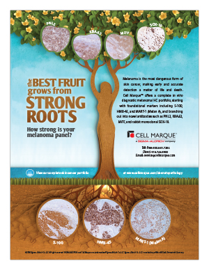
Anti-MART-1 (M2-7C10) demonstrates strong cytoplasmic reaction in this metastatic melanoma.


MART-1 (Melan A) (M2-7C10) Mouse Monoclonal Antibody
Specialties: Dermatopathology
Updated: 2017-05-24 08:46:09
MART-1 (also known as Melan A) is a melanocyte differentiation antigen.1-2 MART-1 is a transmembrane protein present in melanocytes of normal skin, retina, nevi, and most melanomas. MART-1 is a very useful marker for identifying metastatic melanomas.3-11
- Kawakam Y, et al. Cloning of the gene coding for a shared human melanoma antigen recognized by autologous T cells infiltrating into tumor. Proc. Natl. Acad. Sci. U S A. 1994; 91: 3515-19.
- Couli PG, et al. A new gene coding for a differentiation antigen recognized by autologous cytolytic T lymphocytes on HLA-A2 melanomas. J Exp Med. 1994; 180: 35-42.
- Kageshita T, et al. Differential expression of MART-1 in primary and metastatic melanoma lesions. J Immunother. 1997; 20:460-5.
- Fetsch PA, et al. Melanoma-associated antigen recognized by T cells (MART-1): the advent of a preferred immunocytochemical antibody for the diagnosis of metastatic malignant melanoma with fine-needle aspiration. Cancer. 1999; 87:37-42.
- Bergman R, et al. A comparative immunohistochemical study of MART-1 expression in Spitz nevi, ordinary melanocytic nevi, and malignant melanomas. J Am Acad Dermatol. 2000; 42:496-500.
- Orosz Z. Melan-A/Mart-1 expression in various melanocytic lesions and in non-melanocytic soft tissue tumours. Histopathology. 1999; 34:517-25.
- Yaziji H, et al. Immunohistochemical markers of melanocytic tumors. Int J Surg Pathol. 2003; 11:11-5.
- Suchak R, et al. Evaluation of the role of routine melan-A immunohistochemistry for exclusion of microinvasion in 120 cases of lentigo maligna. Am J Dermatopathol. 2014; 36:387-91.
- Helm K et al. Immunohistochemistry of pigmented actinic keratoses, actinic keratoses, melanomas in situ and solar lentigines with Melan-A. J Cutan Pathol. 2008; 35:931-4.
- Kucher C, et al. Expression of Melan-A and Ki-67 in desmoplastic melanoma and desmoplastic nevi. Am J Dermatopathol. 2004; 26:452-7.
- Nielsen PS, et al. Immunohistochemical double stains against Ki67/MART1 and HMB45/MITF: promising diagnostic tools in melanocytic lesions. Am J Dermatopathol. 2011; 33:361-70.
Specifications
- Reactivity: paraffin
- Visualization: cytoplasmic
- Control: melanoma
- Dilution Range: 1:100-1:500*
Package Inserts
IFU
- IVD Rev. 5.0 (CMC28129050)
Have a different keycode?
Click Here
Learn how to obtain your SDS
Ordering Information
For in vitro diagnostic (IVD) use in USA
| 25 mL predilute | 281M-90 |
| 0.1 mL concentrate | 281M-94 |
| 0.5 mL concentrate | 281M-95 |
| 1 mL concentrate | 281M-96 |
| 1 mL predilute | 281M-97 |
| 7 mL predilute | 281M-98 |
For in vitro diagnostic (IVD) use in Canada
| 25 mL predilute | 281M-90 |
| 0.1 mL concentrate | 281M-94 |
| 0.5 mL concentrate | 281M-95 |
| 1 mL concentrate | 281M-96 |
| 1 mL predilute | 281M-97 |
| 7 mL predilute | 281M-98 |
For in vitro diagnostic (IVD) use in Europe
| 25 mL predilute | 281M-90 |
| 0.1 mL concentrate | 281M-94 |
| 0.5 mL concentrate | 281M-95 |
| 1 mL concentrate | 281M-96 |
| 1 mL predilute | 281M-97 |
| 7 mL predilute | 281M-98 |
For research use only (RUO) in Japan
| 25 mL predilute | 281M-90-RUO |
| 0.1 mL concentrate | 281M-94-RUO |
| 0.5 mL concentrate | 281M-95-RUO |
| 1 mL concentrate | 281M-96-RUO |
| 1 mL predilute | 281M-97-RUO |
| 7 mL predilute | 281M-98-RUO |
To request information on this product in additional countries, please click the button below.