


IgD (EP173) Rabbit Monoclonal Antibody
Specialties: Hematopathology
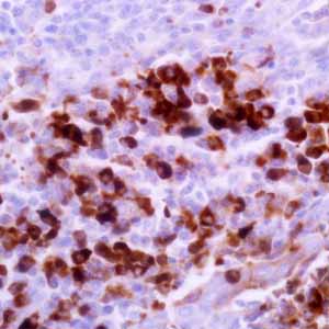
The monoclonal antibody against IgD reacts with immunoglobin D delta chains. In tonsil and lymph node, immunohistochemical staining for IgD immunoglobulin heavy chain is usually used to highlight the tonsil and nodal architecture since the IgD antibody stains mantle zone cells in secondary follicles and mantle cells in primary follicles.1 It has been reported that IgD can be detected in the surface/cytoplasm of neoplastic cells of common small B lymphoid cell lymphomas, such as small lymphocytic lymphoma, mantle cell lymphoma, marginal zone lymphoma (especially splenic marginal zone lymphoma), and follicular lymphoma.1-2 IgD expression in L & P cells of nodular lymphocyte predominant Hodkin lymphoma has been seen in subsets of cases (27% to 71.4%).1-3 The IgD positive L & P cells are usually located in the extrafollicular area with a relatively T-cell-rich background1,3 IgD expression is rarely seen in T-cell rich B-cell lymphoma. Studies have demonstrated that Reed-Sternberg cells of classic Hodgkin lymphoma were negative for IgD1,3 IgD multiple myeloma is a rare bone marrow plasma cell dyscrasia and can be identified by the IgD antibody, especially when a dry tap is encountered.4
- Prakash S, et al. IgD positive L&H cells identify a unique subset of nodular lymphocyte predominant Hodgkin lymphoma. Am J Surg Pathol. 2006; 30:585-92.
- Sohani A, et al. Nodular lymphocyte-predominant Hodgkin lymphoma with atypical T cells: a morphologic variant mimicking peripheral T-cell lymphoma. Am J surg Pathol. 2011; 35:1666-78.
- Kluin, PM, et al. Paediatric nodal marginal zone B-cell lymphadenopathy of the neck: a Haemophilus influenzae-driven immune disorder? J Pathol. 2015; 236:302-14.
- Pandey S, et al. Unusual myelomas: a review of IgD and IgE variants. Oncology (Williston Park). 2013; 27:798-803.
Specifications
- Reactivity: paraffin
- Visualization: cytoplasmic
- Control: tonsil
- Dilution Range: 1:25-1:100*
Package Inserts
IFU
- IVD Rev. 1.0 (CMC26831010)
Have a different keycode?
Click Here
Learn how to obtain your SDS
Ordering Information
For in vitro diagnostic (IVD) use in USA
| 0.1 mL concentrate | 268R-14 |
| 0.5 mL concentrate | 268R-15 |
| 1 mL concentrate | 268R-16 |
| 1 mL predilute | 268R-17 |
| 7 mL predilute | 268R-18 |
For in vitro diagnostic (IVD) use in Canada
| 0.1 mL concentrate | 268R-14 |
| 0.5 mL concentrate | 268R-15 |
| 1 mL concentrate | 268R-16 |
| 1 mL predilute | 268R-17 |
| 7 mL predilute | 268R-18 |
For in vitro diagnostic (IVD) use in Europe
| 0.1 mL concentrate | 268R-14 |
| 0.5 mL concentrate | 268R-15 |
| 1 mL concentrate | 268R-16 |
| 1 mL predilute | 268R-17 |
| 7 mL predilute | 268R-18 |
For research use only (RUO) in Japan
| 0.1 mL concentrate | 268R-14-RUO |
| 0.5 mL concentrate | 268R-15-RUO |
| 1 mL concentrate | 268R-16-RUO |
| 1 mL predilute | 268R-17-RUO |
| 7 mL predilute | 268R-18-RUO |
To request information on this product in additional countries, please click the button below.