SOX-10 (EP268) Rabbit Monoclonal Primary Antibody
Specialties: Dermatopathology
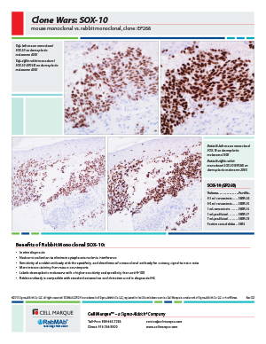
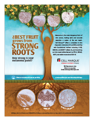
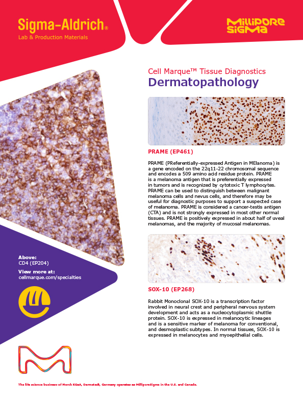
Detection of SOX-10 by immunohistochemistry (IHC) with SOX-10 (EP268) Rabbit Monoclonal Primary Antibody may be used to aid in the diagnosis of melanoma.
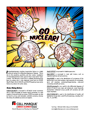
SOX-10 is a member of the SOX (SRY-related HMG-box) family of transcription factors, which are involved in the regulation of embryonic development. SOX-10 plays a role in neural crest development during embryogenesis. Neural crest cells arise from the embryonic ectoderm and give rise to diverse cell types including melanocytes, smooth muscle, peripheral and enteric neurons, and glia.1 SOX-10 is expressed normally in melanocytes, glial cells, and Schwann cells, as well as myoepithelial cells and some salivary gland cells.2-3 SOX-10 expression is also seen in most melanomas.2-4 SOX-10 immunohistochemistry shows a nuclear pattern of staining.
- Rehberg, Stephan et al. "Sox10 is an active nucleocytoplasmic shuttle protein, and shuttling is crucial for Sox10-mediated transactivation." Molecular and cellular biology vol. 22,16 (2002): 5826-34.
- Nonaka, Daisuke et al. "Sox10: a pan-schwannian and melanocytic marker." The American journal of surgical pathology vol. 32,9 (2008): 1291-8.
- Mohamed, Amr et al. "SOX10 expression in malignant melanoma, carcinoma, and normal tissues." Applied immunohistochemistry & molecular morphology : AIMM vol. 21,6 (2013): 506-10.
- Tacha, David et al. "A newly developed mouse monoclonal SOX10 antibody is a highly sensitive and specific marker for malignant melanoma, including spindle cell and desmoplastic melanomas." Archives of pathology & laboratory medicine vol. 139,4 (2015): 530-6.
- Nadji M, Morales AR. Immunoperoxidase, part I: the techniques and its pitfalls. Lab Med 1983;14:767
- Omata M, Liew CT, Ashcavai M, Peters RL. Nonimmunologic binding of horseradish peroxidase to hepatitis B surface antigen. A possible source of error in immunohistochemistry. Am J Clin Pathol. 1980 May; 73(5):626-32. doi: 10.1093/ajcp/73.5.626. PMID: 6155065.
Specifications
- Reactivity: paraffin
- Control: Melanoma (Nuclear); Skin Melanocytes (Nuclear)
- Dilution Range: 1:50-1:200 *
Package Inserts
IFU
- IVD Rev. 5.0 (CMC38331050)
Have a different keycode?
Click Here
Learn how to obtain your SDS
Ordering Information
For in vitro diagnostic (IVD) use in USA
| 25.0 mL predilute ready-to-use | 383R-10 |
| 0.1 mL concentrate | 383R-14 |
| 0.5 mL concentrate | 383R-15 |
| 1.0 mL concentrate | 383R-16 |
| 1.0 mL predilute ready-to-use | 383R-17 |
| 7.0 mL predilute ready-to-use | 383R-18 |
For in vitro diagnostic (IVD) use in Canada
| 25.0 mL predilute ready-to-use | 383R-10 |
| 0.1 mL concentrate | 383R-14 |
| 0.5 mL concentrate | 383R-15 |
| 1.0 mL concentrate | 383R-16 |
| 1.0 mL predilute ready-to-use | 383R-17 |
| 7.0 mL predilute ready-to-use | 383R-18 |
For in vitro diagnostic (IVD) use in Europe
| 25.0 mL predilute ready-to-use | 383R-10 |
| 0.1 mL concentrate | 383R-14 |
| 0.5 mL concentrate | 383R-15 |
| 1.0 mL concentrate | 383R-16 |
| 1.0 mL predilute ready-to-use | 383R-17 |
| 7.0 mL predilute ready-to-use | 383R-18 |
For research use only (RUO) in Japan
| 25.0 mL predilute ready-to-use (RUO) | 383R-10-RUO |
| 0.1 mL concentrate (RUO) | 383R-14-RUO |
| 0.5 mL concentrate (RUO) | 383R-15-RUO |
| 1.0 mL concentrate (RUO) | 383R-16-RUO |
| 1.0 mL predilute ready-to-use (RUO) | 383R-17-RUO |
| 7.0 mL predilute ready-to-use (RUO) | 383R-18-RUO |
To request information on this product in additional countries, please click the button below.