GATA3 (L50-823) Mouse Monoclonal Antibody
Specialties: Anatomic Pathology Breast/Gynecological Pathology Genitourinary (GU) Pathology
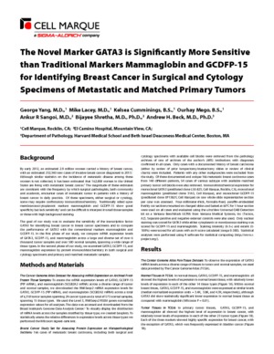
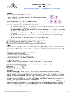
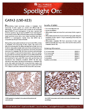
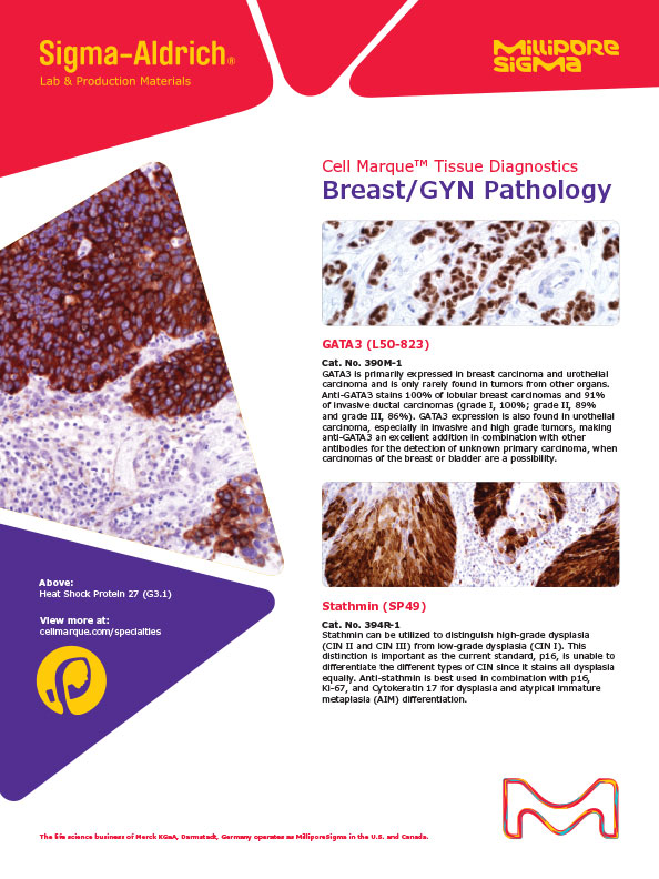
Detection of GATA 3 by immunohistochemistry (IHC) with GATA3 (L50-823) Mouse Monoclonal Antibody may be used to aid in the diagnosis of breast and urothelial carcinomas.
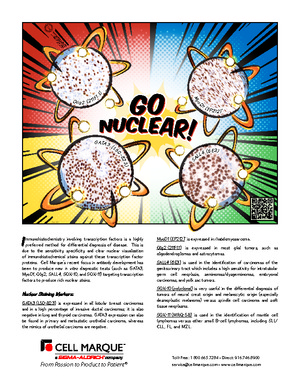
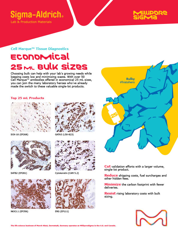
GATA3, or GATA binding protein 3, belongs to the GATA family of tissue specific transcription factors, which play an important role in cell proliferation, development, and differentiation. GATA3 is involved in the differentiation of several tissue types including breast, kidney, and bladder urothelium.1-4 GATA3 expression is seen normally in breast tissue, renal distal tubules, and bladder urothelium.2 GATA3 expression is also found in breast carcinomas and urothelial carcinomas.1-2 Immunohistochemistry for GATA3 shows a nuclear pattern of staining.
- Higgins, John P T et al. "Placental S100 (S100P) and GATA3: markers for transitional epithelium and urothelial carcinoma discovered by complementary DNA microarray." The American journal of surgical pathology vol. 31,5 (2007): 673-80.
- Liu, Haiyan et al. "Immunohistochemical evaluation of GATA3 expression in tumors and normal tissues: a useful immunomarker for breast and urothelial carcinomas." American journal of clinical pathology vol. 138,1 (2012): 57-64.
- Joulin, V et al. "A T-cell specific TCR delta DNA binding protein is a member of the human GATA family." The EMBO journal vol. 10,7 (1991): 1809-16.
- Labastie, M C et al. "Structure and expression of the human GATA3 gene." Genomics vol. 21,1 (1994): 1-6.
- Nadji M, Morales AR. Immunoperoxidase, part I: the techniques and its pitfalls. Lab Med 1983;14:767.
- Omata M, Liew CT, Ashcavai M, Peters RL. Nonimmunologic binding of horseradish peroxidase to hepatitis B surface antigen. A possible source of error in immunohistochemistry. Am J Clin Pathol. 1980 May; 73(5):626-32. doi: 10.1093/ajcp/73.5.626. PMID: 6155065.
Specifications
- Reactivity: paraffin
- Control: Breast carcinoma (Nuclear); Urothelial carcinoma (Nuclear)
- Dilution Range: 1:100-1:500 *
Package Inserts
IFU
- IVD Rev. 5.0 (CMC39021050)
Have a different keycode?
Click Here
Learn how to obtain your SDS
Ordering Information
For in vitro diagnostic (IVD) use in USA
| 25.0 mL predilute ready-to-use | 390M-10 |
| 0.1 mL concentrate | 390M-14 |
| 0.5 mL concentrate | 390M-15 |
| 1.0 mL concentrate | 390M-16 |
| 1.0 mL predilute ready-to-use | 390M-17 |
| 7.0 mL predilute ready-to-use | 390M-18 |
For in vitro diagnostic (IVD) use in Canada
| 25.0 mL predilute ready-to-use | 390M-10 |
| 0.1 mL concentrate | 390M-14 |
| 0.5 mL concentrate | 390M-15 |
| 1.0 mL concentrate | 390M-16 |
| 1.0 mL predilute ready-to-use | 390M-17 |
| 7.0 mL predilute ready-to-use | 390M-18 |
For in vitro diagnostic (IVD) use in Europe
| 25.0 mL predilute ready-to-use | 390M-10 |
| 0.1 mL concentrate | 390M-14 |
| 0.5 mL concentrate | 390M-15 |
| 1.0 mL concentrate | 390M-16 |
| 1.0 mL predilute ready-to-use | 390M-17 |
| 7.0 mL predilute ready-to-use | 390M-18 |
For research use only (RUO) in Japan
| 25.0 mL predilute ready-to-use (RUO) | 390M-10-RUO |
| 0.1 mL concentrate (RUO) | 390M-14-RUO |
| 0.5 mL concentrate (RUO) | 390M-15-RUO |
| 1.0 mL concentrate (RUO) | 390M-16-RUO |
| 1.0 mL predilute ready-to-use (RUO) | 390M-17-RUO |
| 7.0 mL predilute ready-to-use (RUO) | 390M-18-RUO |
To request information on this product in additional countries, please click the button below.