Best IHC Staining Practices
Blocking

Time: 5–15 minutes
Tech Tip: Most blocking is performed prior to primary antibody incubation. In some cases however, a blocking agent may damage or destroy a target epitope. In these instances, it is recommended that blocking is performed after primary antibody incubation.
Insufficient blocking can result in:
Background staining unblocked endogenous tissue elements interact non-specifically with primary antibody, detection, and/or chromogen.
Applying a blocking agent at the wrong time can result in:
No staining blocking agent damages targeted epitope leaving the epitope unrecognizable by the primary antibody; epitope-antibody binding will not occur.
Type:
- Endogenous elements:
Avidin-Biotin block biotin
Peroxide block peroxidase
Alkaline Phosphatase block (Levamisole) phosphatase - Background/Protein block
Tech Tip: Liver, kidney, brain, and spleen contain the highest levels of endogenous biotin. It is important to use an avidin-biotin (A/B) block if utilizing a biotin based detection system.
No blocking or use of an incorrect blocking agent can result in:
Background staining unblocked endogenous tissue elements interact non-specifically with primary antibody, detection, and/or chromogen.
Primary Antibody

Time: 10–60 minutes
Concentration: Variable
Tech Tip: When optimizing a concentrated antibody, it is best to try a minimum of three dilutions; one dilution at the beginning of the manufacturer’s recommended range, one dilution in the middle, and one dilution at the end of the range. Running several dilutions will help account for differences in detection and overall protocol as well as pathologist preference.
If the primary antibody is too diluted or the incubation time is too short, the following may occur:
Weak staining minimal epitope-antibody binding; there is not enough antibody in the solution or the primary antibodies do not have enough time to locate and bind to their targets.
If the primary antibody is too concentrated or the incubation time is too long, the following may occur:
Background staining excess antibody in the solution can bind non-specifically to various sites within the tissue specimen.
Source:
- Polyclonal vs. Monoclonal vs. Rabbit Monoclonal
- Ascites vs. Supernatant
Tech Tip: Polyclonals are a collection of antibody clones that bind to multiple epitopes on a target antigen. Monoclonals are a single antibody clone that binds to one epitope on a target antigen.
Polyclonal and ascites derived primary antibodies can be prone to:
Background staining contain additional immunoglobulins that can non-specifically bind to various elements within a tissue specimen.
Monoclonal and supernatant derived primary antibodies can be prone to:
Weak or No staining target a single epitope; destruction of that epitope during the retrieval or blocking process can minimize binding potential.
Tech Tip: The development of rabbit monoclonal antibodies can yield staining performance utilizing "the best of both worlds". Rabbit monoclonals have the specificity of a mouse monoclonal and the sensitivity of a rabbit polyclonal to enhance single target intensity while minimizing background staining.
Detection
Time: 15–45 minutes
Concentration: Variable
Tech Tip: Most universal detection systems contain anti-mouse/anti-rabbit secondary antibodies.
If the detection system is too dilute or the incubation time too short, the following may occur:
Weak staining minimal detection component binding; overall weak signal.
If the detection system is too concentrated or the incubation time too long, the following may occur:
Background staining excess detection components bind non-specifically to various sites within the tissue specimen.
Handling and Storage: refrigerated at 2–8°C
Tech Tip: It is important to follow the manufacturer’s detection protocol recommendation. Most 2-step detections will not work if the components are applied in the wrong order.
If the detection system is not stored according to manufacturer’s recommendation, the following may occur:
Weak or No staining contamination, degradation of detection components. If the integrity of the detection components is compromised, proper binding can be negatively impacted and overall staining diminished.
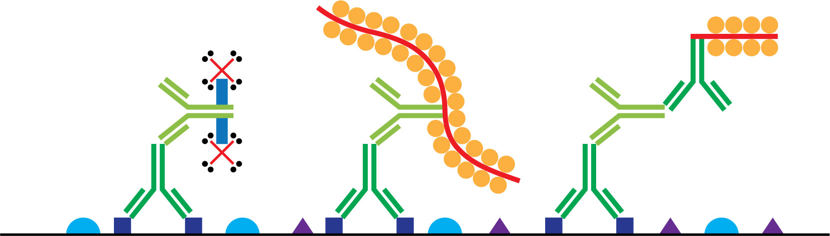
Type and Compatibility: LSAB (Link-Label) vs. 1-step polymer vs. 2-step polymer
Weak staining low detection sensitivity; LSAB (Link-Label) detection systems have fewer color inducing enzymes compared to the much larger polymer systems. Since there is limited opportunity for chromogen-enzyme interaction, the overall signal (staining intensity) is reduced.
Background staining high detection sensitivity; 2-step polymers utilize an additional linking or secondary antibody step. Use of secondary and tertiary antibodies increases the opportunity for non-specific binding to occur.
Tech Tip: Although one-step polymer detection systems have more chromogen binding sites, the bulky polymer molecules are susceptible to steric hindrance, decreasing overall sensitivity.
Chromogen
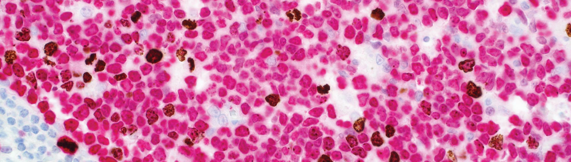
Double Stain Photo Details: DAB (Brown): Phosphohistone H3 (PHH3) (polyclonal); Permanent Red: Ki-67 (SP6)
Time: 1–10 minutes
Concentration: Variable
Tech Tip: Sticky substances/cells, like mucus/mucins, will grab onto excess chromogen resulting in non-specific staining. A short enzyme incubation during or after pretreatment can minimize this non-specific binding.
If the chromogen is too diluted or the incubation time too short, the following may occur:
Weak staining minimal enzyme-chromogen color-producing reaction.
If the chromogen is too concentrated or the incubation time too long, the following may occur:
Background staining chromogen will bind non-specifically. Chromogen is also easily trapped in folds/artifacts created during tissue processing.
Type and Compatibility:
| Horseradish Peroxidase (HRP) | Alkaline Phosphatase (AP) |
|---|---|
| DAB (Brown) | Permanent Red |
| AEC (Red) | Permanent Magenta |
Tech Tip: DAB and AEC will undergo an oxidation-reduction reaction when exposed to HRP; a colored precipitate results. Permanent red and Permanent magenta will lose a phosphate group when exposed to AP; a colored precipitate results.
If an enzyme is used with an incompatible chromogen (AP with DAB for example), the following may occur:
No staining color changing chemical reaction will not occur.
Handling and Storage: refrigerated at 2–8°C
Tech Tip: All chromogens are light sensitive and must be stored in opaque containers.
If the chromogen is not stored according to the manufacturer’s recommendations, the following may occur:
Weak or No staining contamination or oxidation can compromise the integrity of the chromogen.