Best IHC Staining Practices
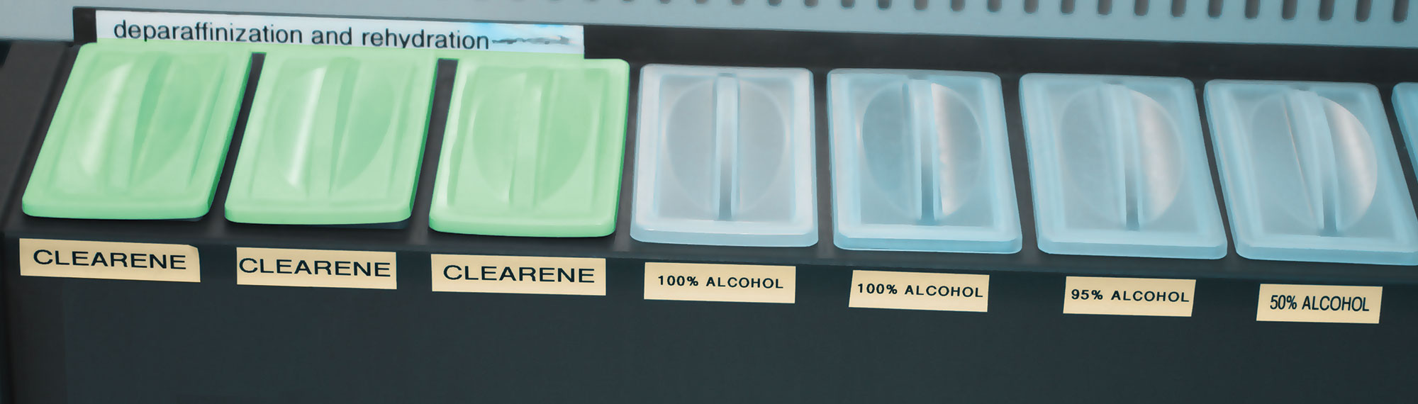
Deparaffinization & Rehydration
Time:
- Deparaffinization: up to 45 minutes; protocols vary
- Rehydration: up to 20 minutes; protocols vary
Tech Tip: Deparaffinization and rehydration protocols can vary depending on the type/strength of reagents used as well as the intensity of the epitope retrieval procedure.
Insufficient deparaffinization can result in:
- Weak or No staining inadequate paraffin removal. If paraffin is not removed, epitopes will not be fully exposed leaving them unrecognizable by the primary antibody.
- Background staining paraffin traps excess chromogen. Paraffin that is not removed will also be evident under the microscope and can be distracting to pathologists.
Type:
- Deparaffinization: Xylene or substitute
- Rehydration: Graded alcohol (100%, 95%, 50%)
Tech Tip: Less toxic xylene substitutes (e.g. Clearene™) are not as potent and will require slightly longer protocols.
Weak or No staining inappropriate or ineffective reagent(s) are used; target epitopes are not retrieved and primary antibody cannot bind.
Weak or No staining reagents become saturated; reagents like xylene and alcohol need to be changed based on usage. If not changed regularly, potency will be lost and deparaffinization/rehydration can become ineffective; paraffin is not removed and the target epitope is still blocked.
Epitope Retrieval
Time: 10–60 minutes; protocols vary
- HIER (heat induced epitope retrieval) 30–60 minutes
- EIER (enzyme induced epitope retrieval) 10–20 minutes
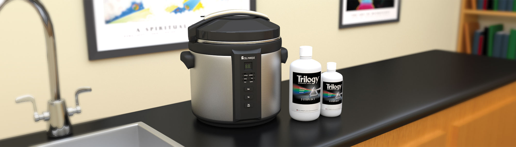
Tech Tip: Protocols vary depending on the type of retrieval (HIER or EIER) and the instrument being used. Pressure cookers allow for the shortest HIER protocols since most will achieve high temperatures of ~120°C.
Insufficient epitope unmasking can result in:
Weak or No staining epitopes are not fully exposed leaving them unrecognizable by the primary antibody; epitope-antibody binding will not occur.
Excessive epitope unmasking can result in:
Background staining tissue will become “over-exposed” with primary antibody and chromogen binding non-specifically to various cellular elements within the over-exposed tissue.
Type:
- HIER (heat induced epitope retrieval)
- EIER (enzyme induced epitope retrieval)
Tech Tip: It is important to follow a manufacturer’s epitope unmasking recommendation. False positives/negatives can occur if not followed correctly.
Use of an incompatible epitope unmasking method can result in:
Weak or No staining some epitopes are better unmasked with either EIER or HIER. If the preferred method is not followed, inadequate epitope retrieval can occur resulting in the primary antibody being unable to bind.
- HIER: Citrate buffer, pH 5–6 and EDTA buffer, pH 8–9
- EIER (Protease): Proteinase K, Pronase, Pepsin, Trypsin
- Equipment: pressure cooker, steamer, waterbath, microwave
Tech Tip: The majority of IHC antibodies on the market will perform optimally using HIER with an EDTA or TRIS buffer (pH 8–9).
Use of an incompatible epitope retrieval reagent can result in:
Weak or No staining inadequate epitope retrieval will occur resulting in the antibody being unable to bind to its target.